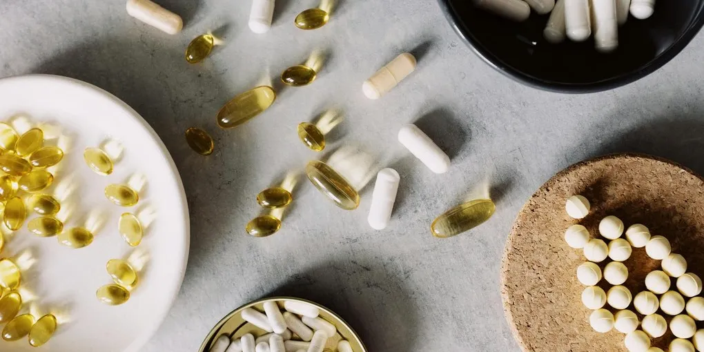
CoQ10 Ubiquinol (Jarrow QH-absorb)
Best Mitochondrial Supplement — Editor's PickForm: Ubiquinol (reduced active form)
$35–60 / 60 softgels
Quick Comparison
| Product | Key Specs | Price Range | Buy |
|---|---|---|---|
| CoQ10 Ubiquinol (Jarrow QH-absorb) Best Mitochondrial Supplement — Editor's Pick |
| $35–60 / 60 softgels | Check Price on Amazon |
| NMN Supplement (Alive by Nature) Best NAD+ Precursor |
| $40–70 / 60 capsules | Check Price on Amazon |
| R-Alpha Lipoic Acid (Jarrow R-ALA) Best Mitochondrial Antioxidant |
| $25–40 / 60 capsules | Check Price on Amazon |
Contains affiliate links — we may earn a small commission at no extra cost to you.
Mitochondrial Health: How to Optimize Your Cellular Energy Production
Mitochondria are central to nearly every disease of aging. Alzheimer’s disease, type 2 diabetes, heart failure, Parkinson’s disease, sarcopenia, and cancer all involve mitochondrial dysfunction as a contributing mechanism. They are not peripheral — they are at the intersection of energy metabolism, inflammation, oxidative stress, and cellular death.
This guide covers what mitochondria actually do, the mechanisms of mitochondrial decline, and the evidence-based interventions that protect and improve mitochondrial function — organized from highest to lowest impact.
Mitochondria: Function and Structure
Each human cell contains hundreds to thousands of mitochondria (neurons and heart cells among the most mitochondria-dense). They are double-membraned organelles with their own circular DNA (mtDNA) — an evolutionary remnant of the alpha-proteobacterial ancestor that became endosymbiotic in eukaryotic cells approximately 1.5 billion years ago.
Core functions:
ATP production via oxidative phosphorylation (OXPHOS): The electron transport chain (ETC) comprises five protein complexes (I–V) embedded in the inner mitochondrial membrane. Electrons derived from NADH and FADH2 (produced in the TCA cycle from carbohydrates, fats, and amino acids) are passed down the chain to molecular oxygen (the terminal electron acceptor). The proton gradient generated drives ATP synthase (Complex V) to produce ATP. The brain, heart, and skeletal muscle are the most energetically demanding organs — their function is tightly coupled to mitochondrial output.
ROS production and signaling: A small percentage (~0.1–2%) of electrons “leak” from the ETC and combine with oxygen to form superoxide, the precursor to hydrogen peroxide and other reactive oxygen species. At physiological concentrations, ROS are redox signaling molecules that activate adaptive responses (including NRF2, the master antioxidant transcription factor). At excessive concentrations — produced by damaged or overloaded mitochondria — ROS cause lipid peroxidation, protein carbonylation, and mtDNA damage.
Calcium homeostasis: Mitochondria buffer cytosolic calcium, integrating metabolic demand with cellular signaling. Dysregulation of mitochondrial calcium handling is implicated in ischemia-reperfusion injury and neurodegeneration.
Apoptosis regulation: The intrinsic apoptosis pathway is controlled by the permeability of the outer mitochondrial membrane. Release of cytochrome c into the cytoplasm triggers caspase cascades. Mitochondria are the gatekeepers of programmed cell death — essential for development and immune function, pathological when dysregulated.
Mitochondrial Decline: The Aging Connection
Mitochondrial dysfunction accumulates with aging through multiple compounding mechanisms:
-
mtDNA mutation accumulation: mtDNA lacks the histone protection of nuclear DNA and is in close proximity to the ETC — the primary source of ROS. Mutation rates are 10–17x higher than nuclear DNA. Mutations in ETC subunits reduce Complex I–IV efficiency, increasing electron leak and ROS production in a vicious cycle.
-
Reduced mitochondrial biogenesis: PGC-1α expression declines with age and sedentary behavior. Fewer new mitochondria are produced to replace dysfunctional ones.
-
Impaired mitophagy: The cellular machinery for identifying and destroying dysfunctional mitochondria (PINK1/Parkin pathway) becomes less efficient with age. Damaged mitochondria accumulate and propagate their dysfunction to daughter cells.
-
NAD+ decline: NAD+ is a critical redox cofactor and substrate for sirtuins (SIRT1, SIRT3) and PARP enzymes. NAD+ levels decline 40–60% between young adulthood and middle age in multiple tissues (Yoshino et al., 2011; PMID: 21884979). NAD+ depletion reduces SIRT3 activity, which normally deacetylates and activates ETC components.
-
Reduced mitochondrial fission/fusion dynamics: Mitochondria constantly merge (fusion) and divide (fission) to mix their contents, segregate damaged components, and optimize ATP output. This quality control process is impaired with aging, obesity, and neurodegeneration.
The Highest-Impact Interventions
1. Exercise — The Primary Biogenesis Signal
Exercise is irreplaceable. No supplement activates mitochondrial biogenesis as powerfully or comprehensively as physical training.
Endurance exercise activates PGC-1α via multiple upstream signals: AMPK (activated by low energy charge), p38 MAPK (activated by contractile stress), and calcium/calmodulin kinase (CaMKII, activated by calcium release). PGC-1α then drives transcription of nuclear-encoded mitochondrial genes and increases mitochondrial density. Hood et al. established that 6 weeks of endurance training increases mitochondrial enzyme activity (markers of mitochondrial content) in skeletal muscle by 30–50%.
High-intensity training (HIIT) activates mitophagy in addition to biogenesis — the combination of clearing damaged mitochondria while stimulating production of new ones is more complete than endurance training alone. HIIT also more powerfully activates PGC-1α acutely, with up to 10-fold PGC-1α mRNA increases following a single HIIT session.
Resistance training increases mitochondrial content in Type II (fast-twitch) muscle fibers — a population often neglected by endurance-only protocols. For older adults, resistance training is particularly critical as Type II fiber atrophy and mitochondrial decline in fast-twitch muscle is a primary driver of age-related functional decline.
Practical minimum: 150 minutes/week moderate aerobic + 2 sessions/week resistance training. For mitochondrial optimization, include 1–2 HIIT sessions per week.
2. Caloric Restriction and Fasting
Caloric restriction (CR) is the most studied and consistent intervention for extending mitochondrial function and lifespan in model organisms. In humans, a 25% CR protocol in the CALERIE trial (Kraus et al., 2019; PMID: 30830035) improved cardiometabolic biomarkers and increased energy expenditure efficiency — suggesting metabolic adaptation toward mitochondrial efficiency.
Caloric restriction activates AMPK and SIRT1, which coordinate PGC-1α activation and improved mitochondrial function.
Intermittent fasting (IF): Time-restricted eating (TRE) and periodic fasting protocols activate similar AMPK/SIRT1 pathways with less sustained caloric restriction. Fasting also stimulates mitophagy — the removal of damaged mitochondria is upregulated during periods of cellular energy deficit. De Cabo & Mattson (2019; PMID: 31881139) reviewed the mechanisms and clinical evidence for IF, noting improvements in metabolic biomarkers, oxidative stress markers, and inflammatory cytokines in multiple human trials.
3. Cold and Heat Hormesis
Cold exposure: Cold activates thermogenesis in brown adipose tissue (BAT), which is densely packed with mitochondria specifically for heat production. Repeated cold exposure increases BAT mass and mitochondrial uncoupling capacity. In skeletal muscle, cold-induced shivering activates AMPK and modest PGC-1α upregulation.
Heat exposure (sauna): Heat stress activates heat shock proteins (HSPs), including HSP70 and HSP27, which stabilize mitochondrial protein complexes, reduce protein aggregation, and maintain ETC efficiency under stress. A Finnish cohort study (Laukkanen et al., 2017; PMID: 28116148) found that frequent sauna use (4–7x/week) was associated with dose-dependent reductions in cardiovascular mortality — consistent with cumulative cardioprotective and mitochondrial protective effects.
Evidence-Based Supplements for Mitochondrial Support
Coenzyme Q10 (CoQ10) / Ubiquinol: CoQ10 is an endogenous component of the ETC (shuttles electrons between Complexes I/II and Complex III) and the primary lipid-phase antioxidant in the inner mitochondrial membrane. Plasma CoQ10 declines with age and is dramatically reduced by statin medications (statins inhibit the mevalonate pathway that produces CoQ10 as well as cholesterol). Ubiquinol (the reduced form) has superior bioavailability over ubiquinone in older adults. Meta-analyses of CoQ10 supplementation show consistent improvement in exercise performance and reduction in oxidative stress biomarkers (Hargreaves & Bhattacharya, 2012). Dose: 100–300 mg ubiquinol/day.
NAD+ Precursors (NMN, NR): NAD+ is the essential substrate for SIRT1 and SIRT3 — sirtuin deacetylases that maintain mitochondrial quality control, biogenesis, and ETC efficiency. Supplementing with NMN (nicotinamide mononucleotide) or NR (nicotinamide riboside) raises systemic and tissue NAD+ levels. Yoshino et al. (2021; PMID: 34449789) showed NMN supplementation in postmenopausal prediabetic women increased muscle NAD+ levels and improved muscle insulin sensitivity. Dose: 250–500 mg NMN or 300–600 mg NR daily.
PQQ (Pyrroloquinoline Quinone): PQQ activates mitochondrial biogenesis independently of PGC-1α through NRF2 and CREB pathways. Nakano et al. (2012; PMID: 22431056) showed 20 mg PQQ/day for 8 weeks significantly increased urinary 8-OHdG (oxidative stress marker) reduction and improved inflammatory markers in a small human trial. Dose: 10–20 mg/day.
Alpha-Lipoic Acid (ALA): ALA is an endogenous cofactor for mitochondrial enzyme complexes (pyruvate dehydrogenase, alpha-ketoglutarate dehydrogenase) and a potent mitochondrial antioxidant that recycles vitamin C, E, and glutathione. R-ALA (the naturally occurring form) is more bioactive than the racemic S+R mix in most supplements. Dose: 200–600 mg R-ALA/day.
Urolithin A: Urolithin A is a gut-microbial metabolite of ellagitannins (from pomegranates, walnuts) that is the most potent dietary activator of mitophagy identified in humans. Andreux et al. (2019; PMID: 30854183) showed oral urolithin A supplementation (500–2000 mg) increased mitophagy biomarkers in muscle and plasma of older adults in a first-in-human RCT. Dose: 500–1000 mg/day of Mitopure® (tested form).
Summary
Mitochondrial health is foundational to energy, performance, metabolic health, and longevity. The highest-impact interventions are exercise (particularly HIIT + endurance combination), caloric appropriateness, intermittent fasting, and quality sleep — all of which activate the core pathways (PGC-1α, AMPK, SIRT1) that drive mitochondrial biogenesis and quality control. Evidence-based supplements (CoQ10/ubiquinol, NAD+ precursors, urolithin A) can support these foundations, particularly in older adults or those with known mitochondrial stressors.
Affiliate disclosure: This article contains Amazon affiliate links. Body Science Review may earn a commission on qualifying purchases at no additional cost to you. This does not influence our editorial conclusions.
AI transparency: This article was researched and drafted with AI assistance and reviewed for factual accuracy against peer-reviewed sources.
Frequently Asked Questions
- Mitochondria are best known for producing ATP (adenosine triphosphate) — the primary cellular energy currency — via oxidative phosphorylation. But their role is far broader. Mitochondria regulate calcium signaling, apoptosis (programmed cell death), reactive oxygen species (ROS) production and scavenging, thermogenesis in brown adipose tissue, steroid hormone synthesis, and immune signaling. They are sensors of cellular stress that coordinate the cell's response to metabolic challenges.
- Mitochondrial biogenesis is the process by which cells increase their mitochondrial number and density. It is regulated primarily by PGC-1α (peroxisome proliferator-activated receptor gamma coactivator 1-alpha), a transcriptional coactivator activated by exercise, caloric restriction, cold exposure, and certain compounds. More mitochondria per cell means greater capacity for aerobic energy production, better endurance performance, and stronger metabolic health.
- Mitochondrial dysfunction — impaired ATP production, excessive ROS generation, and failure of quality control mechanisms — is driven by sedentary lifestyle, chronic caloric excess, chronic sleep deprivation, environmental toxin exposure (heavy metals, pesticides), aging-related decline in mitochondrial quality control, and genetic mitochondrial diseases (rare). The most prevalent and reversible causes are physical inactivity and metabolic excess.
- Supplements can support mitochondrial function and reduce oxidative damage, but they cannot replace the foundational interventions — exercise, caloric appropriateness, and sleep. CoQ10 and MitoQ have the strongest evidence for supporting mitochondrial ETC function. NAD+ precursors (NMN, NR) support the redox cofactor supply. PQQ and alpha-lipoic acid show antioxidant effects at mitochondria. None replace the biogenesis signal provided by exercise.
- Mitophagy is the selective autophagy of damaged or dysfunctional mitochondria. It is the cellular quality control mechanism that removes mitochondria with compromised membrane potential before they accumulate ROS and trigger apoptosis. Healthy mitophagy declines with age and sedentary behavior. Exercise, fasting, and urolithin A are the most evidence-supported stimuli for mitophagy in humans.